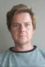
Bob Asselbergh
Microscopy Expert - Research Associate

Science
After obtaining my PhD in 2007 I pursued my growing passion
for microscopy and obtained expertise in several microscopy-related techniques.
I address the challenge of integrating the optimal imaging and analysis
techniques in a biological context to answer questions related to complex
neurological diseases of the central and peripheral nervous system. An
important research goal in the center is to study the pathogenic role of
mutations that are identified by genetic approaches. To this end, we employ
Drosophila, mouse and several cellular model systems to study the molecular and
biological mechanisms of how these mutations cause neurological disease. The
use of microscopy and the establishment of quantitative imaging assays are
providing valuable tools to analyze the complex cell biological disturbances
that result from these mutations. In my function, I provide scientific and
technological microscopy-related support for the different research projects in
our department. Other responsibilities include the maintenance and acquisition
of microscopy equipment (light, fluorescence and confocal microscopes),
training of researchers on the different microscopy systems and in digital imaging,
and establishing networks for microscopy expertise in- and outside VIB and the
university.